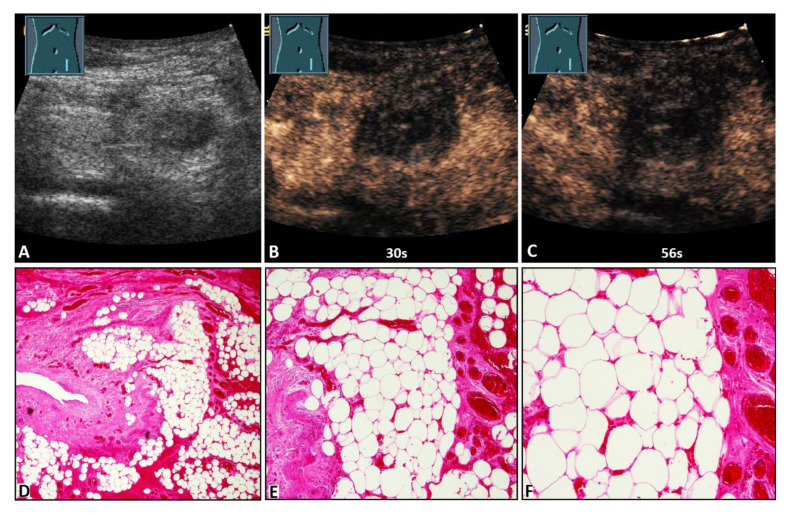
Figure 3.
A 47-year-old female patient with acute peritoneal pain in the left lower abdomen and a palpable inguinal-located tumor. (A) B-mode ultrasound shows a hyperechoic lesion with a central hypoechoic area. (B,C) On contrast-enhanced ultrasound, the lesion shows absent enhancement after 30 s and 56 s, and an omental hernia was diagnosed. (D) Omentum with partial fibrotic consolidation (on the left) and dilated hyperemic capillaries and hemorrhages (4× magnification). (E) Omentum with central necrotic fat tissue (missing nuclei), fibrosis (to the left), and dilated hyperemic capillaries and hemorrhages (on the right) (10× magnification). (F) Omentum with central necrotic fat tissue, and dilated hyperemic capillaries and hemorrhages (20× magnification).

