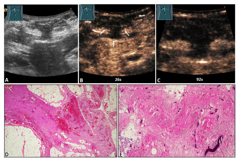
Figure 4.
A 59-year-old male patient with acute peritoneal pain in the umbilical region and a palpable tumor. (A) B-mode ultrasound shows a complex lesion located in the abdominal wall, with visualization of omental hernia content. (B,C) On contrast-enhanced ultrasound, the lesion shows absent enhancement after 26 s (arrows) and 92 s. (D) Omentum with fibrotic strands, including dilated hyperemic capillaries (4× magnification). (E) Fibrotic hernia sac with dilated hyperemic capillaries (10× magnification).

