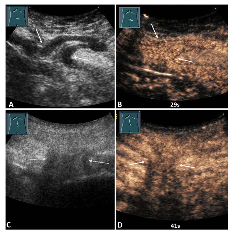
Figure 5.
(A) A 59-year-old male patient with acute pain in the paraumbilical region. B-mode ultrasound shows a hernia orifice (arrows), and the hernia sac shows a fixed bowel structure. (B) On contrast-enhanced ultrasound, the loop of small bowel shows a marked enhancement after 29 s. Surgical evaluation showed no evidence of infarction. No resection of the bowel was performed. (C) A 91-year-old female patient with acute pain in the epigastric region. B-mode ultrasound shows a hernia orifice (arrows), and the hernia sac shows fixed omental tissue. (D) On contrast-enhanced ultrasound, the hernia contents show a marked enhancement after 41 s. Surgical evaluation showed no evidence of infarction. No resection of the omentum was performed.

