. 2022 Feb 17;11:e73220. doi: 10.7554/eLife.73220

Figure 4. Lem8 cleaves Phldb2 in a manner that requires 14-3-3ζ.

(A) Multiple alignments of the self-cleavage site of Lem8 with potential targets in human cells identified by bioinformatic analysis. Identical residues are highlighted in red. (B) Lem8 reduces the protein levels of endogenous Phldb2 in mammalian cells. Lem8 and the indicated mutants were individually expressed in HEK293T cells by transfection. Twenty-four hours after transfection, the samples were resolved by sodium dodecyl sulfate–polyacrylamide gel electrophoresis (SDS–PAGE) and detected by immunoblotting with anti-Phldb2 antibodies. Tubulin was used as a loading control. Results shown were one representative from three independent experiments with similar results. (C) Lem8 cleaves exogenous Phldb2 in mammalian cells. HA and Flag tag were fused to the amino and carboxyl end of Phldb2, respectively, and the double tagged protein was coexpressed in HEK293T cells with Lem8 or each of the mutants. Twenty-four hours after transfection, the samples were resolved by SDS–PAGE and probed by a HA-specific antibody and a Flag-specific antibody, respectively. Tubulin was detected as a loading control. Results shown were one representative from three independent experiments with similar results. (D) Lem8 alters the subcellular distribution of GFP fused to Phldb2. GFP was fused to the amino end of Phldb2 and the protein was coexpressed in HEK293T cells with mCherry-Lem8 or each of the mutants. Twenty-four hours after transfection, cells were fixed and nucleus were stained by Hoechst 33,342. The fluorescence Images of GFP (green), mCherry (red), and Hoechst (blue) were acquired with a Zeiss LSM 880 confocal microscope. The percentage of cells with membrane Phldb2 was calculated in Phldb2- and Lem8-positive cells (right panel). Bar, 10 μm. (E) 14-3-3ζ is required for the cleavage of Phldb2 by Lem8. HA-Phldb2-Flag was expressed in HEK293T cells, immunoprecipitated with a Flag-specific antibody, and eluted with 3× Flag peptides. Purified Phldb2 was incubated with His6-Lem8 or each of the mutants in reactions with or without His6-14-3-3ζ. Total proteins of all samples were resolved with SDS–PAGE, and probed by immunoblotting with a HA-specific antibody, a Flag-specific antibody and a His-specific antibody. Results shown were one representative from three independent experiments with similar results.

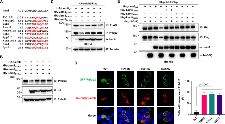
Figure 4.

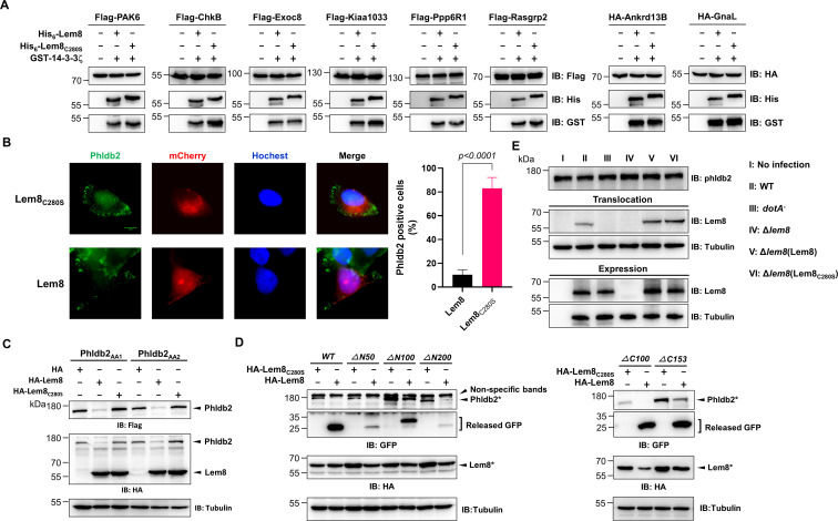
Figure 4—figure supplement 1. Verification of Lem8-mediated cleavage of candidate proteins and its cleavage of phldb2 at multiple sites.

Figure 4—figure supplement 1.

(A) Cleavage of substrate candidates by Lem8. Flag- or HA-tagged Rasgrp2, Pak6, Exoc8, Ankrd13B, Chkb, Ppp6R1, Kiaa1033, Gnal, and Gpr61 expressed in HEK293T cells each was immunoprecipitated with antibodies specific for Flag or HA. Proteins eluted with 3× Flag or HA peptides were incubated with His-Lem8 and His6-14-3-3ζ. Samples resolved with sodium dodecyl sulfate–polyacrylamide gel electrophoresis (SDS–PAGE) were probed by immunoblotting with a Flag- or HA-specific antibody. Note that Flag- or HA-GPR61 did not express so it was not examined. Results shown were one representative from three independent experiments with similar results. (B) Lem8 causes redistribution of Phldb2 in cells. Hela cells were transfected to express the indicated mCherry fusion proteins. Twenty-four hours after transfection, cells were fixed and immunostained with anti-Phldb2 antibodies. The nuclei were stained by Hoechst 33,342. Images were acquired with a Zeiss LSM 880 confocal microscope. Phldb2, green (GFP); Lem8 and its mutants, red (mCherry); nuclei, blue (Hoechst). The percentage of Phldb2-positive cells was calculated in Lem8-positive cells (right panel). Bar, 10 μm. (C) Mutations were introduced into HA-Phldb2-Flag to replace residues Arg1111 and Gln1112 (Phldb2AA1) or Gln1112 and Arg1113 (Phldb2AA2) with alanine, respectively. The two mutants were coexpressed in HEK293T cells with Lem8 or Lem8C280S. Samples were resolved with SDS–PAGE, and probed by immunoblotting with the antibody specific to HA and Flag, respectively. Mutations in the cleavage site of Phldb2 cannot completely prevent its degradation by Lem8. Results shown were one representative from three independent experiments with similar results. (D) Lem8 removes the GFP tag fused to the amino end of Phldb2 deletion mutants. GFP was fused to the amino end of Phldb2 and the indicated truncation mutants. The fusion proteins were individually coexpressed with HA-Lem8 or HA-Lem8C280S in HEK293T cells by transfection. Samples resolved by SDS–PAGE were detected by immunoblotting with GFP-specific antibodies. Results shown were one representative from three independent experiments with similar results. (E) Cleavage of Phldb2 is undetectable during L. pneumophila infection. HEK293T cells transfected to express FcγRII receptor were infected with the indicated bacterial strains. Two hours after infection, the protein levels of Phldb2, as well as the translocation and expression of Lem8, were probed with the appropriate antibodies with Tubulin and ICDH as loading control, respectively. Results shown were one representative from three independent experiments with similar results. Bacterial strains: I, noninfection; II, Lp02 (WT); III, dotA (defective in Dot/Icm); IV, Lp02Δlem8; V, Lp02Δlem8 (pLem8); VI, Lp02Δlem8 (pLem8C280S).