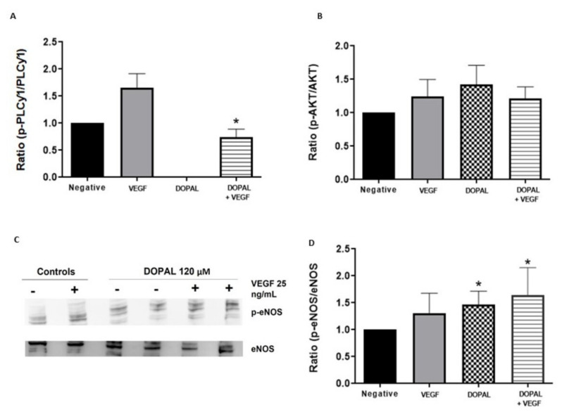
Figure 2.
Effects of DOPAL on PLCγ1, Akt and eNOS. HUVEC cells were treated with DOPAL (120 µM) for 4 h and then stimulated with VEGF (25 ng/mL) for 10 min (A) and 60 min (B–D). Western-blot membranes were incubated with anti PLCγ-1 and anti p-PLCγ-1 (A), anti Akt and anti p-Akt (B) and anti eNOS and anti p-eNOS (C,D) antibodies. Data representation of p-PLCγ-1/PLCγ-1, p-Akt/Akt and p-eNOS/eNOS ratio are displayed as mean ± SD (n = 5). * p < 0.05 against VEGF alone (A) and versus negative control (D).

