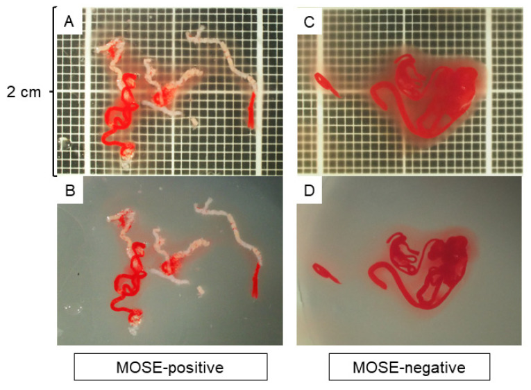
Figure 1.
Macroscopic on-site evaluation (MOSE) using a stereomicroscope. The observation screen was set up with a vertical width of 2 cm with a scale showing 1-mm increments in the background. The quality of the specimen was evaluated without anything in the background, and the positivity of the MOSE results was judged based on the presence of whitish core tissue. (A,B): A specimen judged as MOSE-positive with (A) and without (B) a scale in the background. (C,D): A specimen judged as MOSE-negative with (C) and without (D) a scale in the background.

