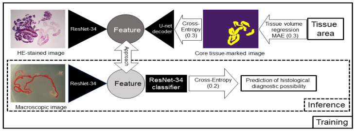
Figure 5.
Schema of the contrastive learning network used for this study. Artificial intelligence (AI) learned the relationships between the haematoxylin and eosin (HE)-stained image and the image with the marked core tissue and detected which pixels corresponded to the tissue. The contrastive learning method was also trained to approach the features of the stereomicroscopic images and HE-stained images, and we investigated whether linking the two images would improve the prediction rate of the AI-based diagnostic method for a positive histological diagnosis. For training, we used the 3 networks surrounded by a square, and for inference, we used only the lower network surrounded by a dotted square. MAE: mean absolute error. The numbers in parentheses represent the weight of each loss.

