Figure 5.

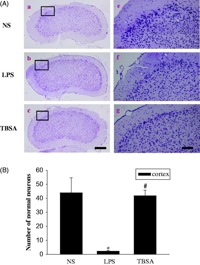
HDAC6 inhibition diminishes neuronal damage in the frontal cortex. Typical images of cresyl violet-stained sections from the frontal cortex of the NS group (a, d), mice injected with LPS (b, e) and mice treated with both LPS and TBSA (c, f). Data were obtained from six independent animals in each experimental group and the results of a typical experiment are presented here. Boxed areas in the left column are shown at higher magnification in the right column. Scale bar in d = 200 μm; Scale bar in h = 20 μm. (B) The cell density in the frontal cortex was calculated. Data were obtained from six independent animals in each experiment group. *p < 0.05 compared with the respective saline group; #p < 0.05 compared with the respective LPS group.
