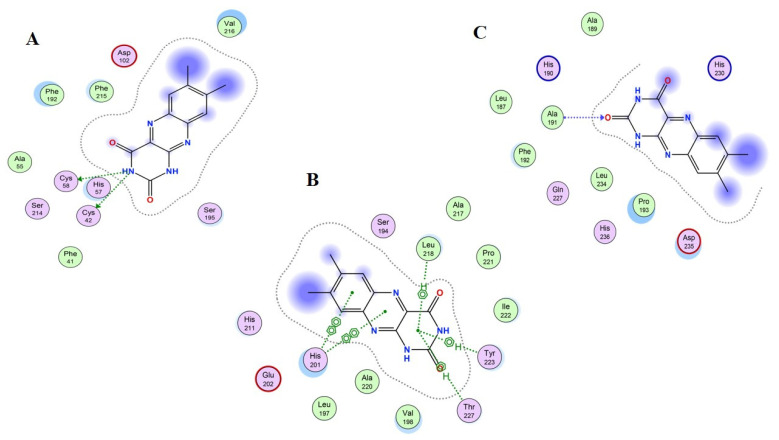
Figure 10.
Schematic representation of Compound 11 “lumichrome” bonded in the active site of selected three proteins (PDB ID: 1H1B (A), 1QIB (B) and 4H1Q (C)). The active binding site shows hydrogen-bonding capacity as donor atoms (purple), and acceptor atoms (green), a two-dimensional plot showing hydrogen bonding interactions and other important hydrophobic interacting residues of the target proteins.

