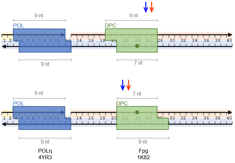
Figure 4.
An arrangement of hPOLη and cross-linked Fpg on DNA with the sizes of the proteins estimated from the structural data. PDB IDs are 1K82 for the structure of Fpg [42] and 4YR3 for the structure of hPOLη [78]. Arrowheads mark the 3′ termini of the oligonucleotides. The green dots indicate the sites of cross-linking. The colored arrows indicate the termination positions of hPOLη while Fpg is cross-linked with the template strand (top panel) or the displaced strand (bottom panel). The blue arrows mark the positions of the last incorporated dNMP, the red arrows show the corresponding positions of the estimated front sides of hPOLη.

