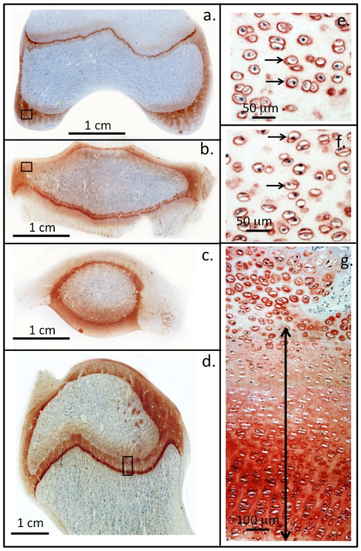
Figure 2.
Perlecan localisation in ovine knee and hip joints. Immunolocalisation of perlecan in cartilaginous tissues of a two-year-old ovine knee femoral condyle (a) and tibial plateau (b), patella (c) and in the humeral head of a hip joint (d). Higher power images demonstrate the pericellular localisation of perlecan (small arrows) around chondrocytes in regions of the femoral (e) and tibial articular cartilages (f) (boxed areas in (a,b)). Perlecan is also present as a gradient throughout the femoral long bone growth plate ECM of the hip in the resting and proliferative zones (double headed arrow) and is prominently expressed pericellularly by the hypertrophic columnar hip chondrocytes located in the bottom of photosegment (g). NovaRED chromogen, perlecan localised with MAb A7L6 to perlecan domain IV. Photo segments (a–g) modified from [9] reproduced under Open Access Creative Commons Attribution 4.0 International licence images © the authors (2010).

