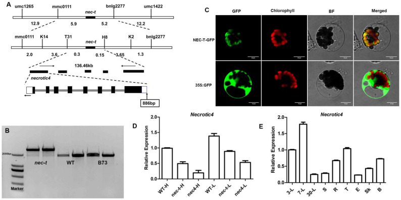
Figure 4.
Map-based cloning and expression analysis of nec-t. (A) Map-based cloning of the nec-t gene. The black region represents the nec-t gene, and the arrow indicates the insertion position [22]. (B) Validation of inserted fragments in nec-t. The amplified fragment of WT and B73 was 2.0 k, and that of nec-t was about 2.9 k. (C) NEC-T protein is co-located with the chloroplast. NEC-T: the coding protein of Nec-t gene; GFP: green fluorescent protein; 35S: 35S promoter; NEC-T-GFP, NEC-T-GFP fusion protein; 35S:GFP: control of GFP protein. bar = 10 μm. (D) Relative expression of the Necrotic4 gene under different temperatures in the leaves of WT, nec-t, and nec4. H, 30 °C; L, 24 °C. (E) Relative expression of the Necrotic4 gene in different tissues. 3-L: Leaves of 3 days after germination; 30-L: Leaves of 30 days after germination; S: stem; R: root; T: tassel; E: ears. Error bars indicating SD were obtained from three biological repeats.

