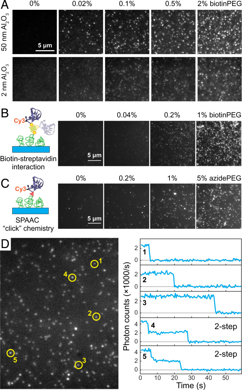
Fig. 2.
Single-molecule characterization of fluorescently labeled biomolecules immobilized on diamond surfaces. (A) Fluorescence images of the immobilized SA-488 molecules for various biotinPEG percentages (0, 0.02, 0.1, 0.5, and 2%) and two different Al2O3 thickness (50 nm, imaged in buffer, and 2 nm, imaged in a refraction index = 1.42 Invitrogen Antifade medium). (B and C) Immobilization of a Cy3-ssDNA on diamond surfaces. This is achieved via either biotin–streptavidin interaction (B) or SPAAC (C). (D) A representative area of single-molecule fluorescence images of in B and the time traces of five selected fluorescence spots.

