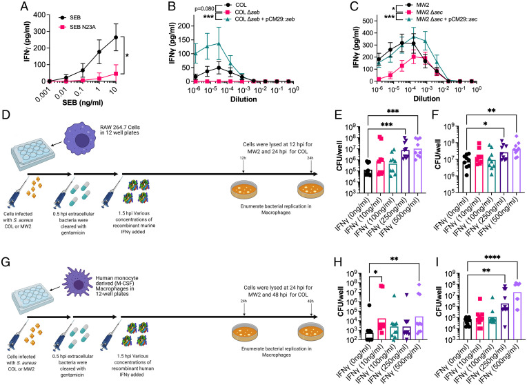
Fig. 7.
SEB and SEC elicit IFN-γ from human cells, and excessive concentration can promote increased extracellular replication of S. aureus in macrophages. (A) IFN-γ production by PBMCs from human blood following stimulation with a titration of SEB or the SEB-N23A proteins. (B and C) IFN-γ production by PBMCs from human blood following stimulation with a titration of supernatant from S. aureus COL (B) or MW2 (C) strains as indicated. Supernatants were taken from cultures grown for 8 h in BHI prior to use in these assays. Data shown (A–C) are mean ± SEM from eight donors. Significant differences were determined from the area under each curve using a paired Friedman test for multiple comparisons (*P < 0.05, ***P < 0.001). (D) Schematic outlining the procedure for intracellular S. aureus replication in RAW 264.7 murine macrophages after dosing with recombinant murine IFN-γ. (E and F) S. aureus recovered from RAW 264.7 cells after incubation at 24 h for strain COL (E) and 12 h for strain MW2 (F) with varying concentrations of recombinant IFN-γ. Each dot represents an individual experiment, and the bar represents the geometric mean for CFUs/well. (G) Schematic outlining the procedure for intracellular S. aureus replication in monocyte-derived human macrophages after dosing with recombinant human IFN-γ. (H and I) S. aureus recovered from human macrophages after incubation at 48 h for strain COL (H) and 24 h for strain MW2 (I) with varying concentrations of recombinant IFN-γ. Each dot represents macrophages from an individual human donor, and the bar represents the geometric mean for CFUs/well. Significant differences between 0 ng/mL of IFN-γ and other concentrations were determined using the Kruskal–Wallis test with the uncorrected Dunn’s test for multiple comparisons (*P < 0.05, **P < 0.01, ****P < 0.0001).

