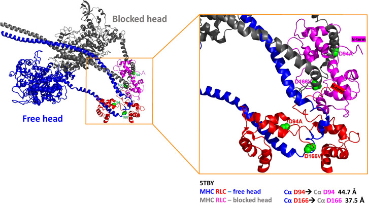
Fig. 5.
The tertiary structure of human β-cardiac heavy meromyosin IHM (Protein Data Bank 5TBY) and PyMOL-derived distances between Cα atoms of D94 and D166 residues positioned on the RLC of the “blocked” head (MHC: gray, RLC: pink) and the “free” head (MHC: dark blue, RLC: red). The side chains of mutations are presented as green spheres, and the N-termini of RLCs are labeled with corresponding colors, pink for the RLC of BH and red for the RLC of FH.

