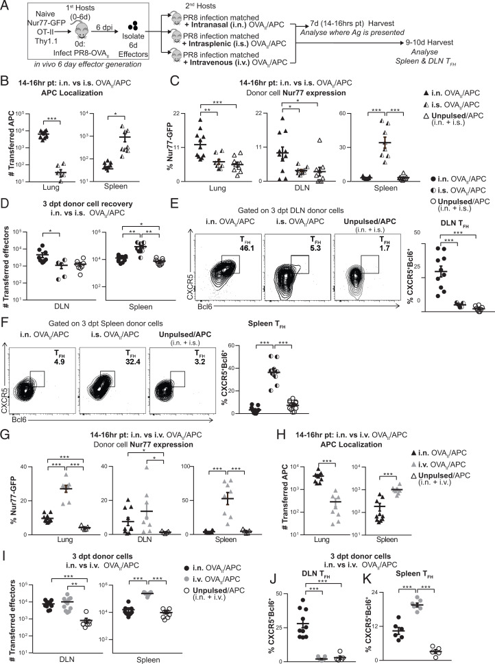
Fig. 4.
Local Ag presentation during the effector phase is required for DLN and spleen TFH as shown by Ag delivery via i.n. vs. i.s vs. i.v. routes. (A) Experimental design: OVAII peptide-pulsed B cells (CD45.1+ or GFP+) were used as APCs and transferred into PR8 infection-matched hosts 6 dpi by either i.n., i.s., or i.v. routes. Unpulsed APCs were transferred both i.n. and i.s., or both i.n. and i.v., as negative controls. In vivo-generated 6-d OT-II.Nur77GFP.Thy1.1+ effectors were transferred by i.v. route. Mice were harvested 14 to 16 h posttransfer (pt), and donor cells were analyzed by flow cytometry. (B and H) Number of transferred APCs. (C and G) Donor cell Nur77GFP expression was analyzed by flow cytometry in the lung, DLN, and spleen. (n = 8 to 11 per group pooled, 3 independent experiments). (D–F, I, and J) Experiment was performed as in A, and mice were euthanized 3 to 4 dpt. (D and I) Total numbers of DLN and spleen donor effectors recovered (n = 5 to 12 per group pooled, 3 to 4 independent experiments). (E and J) DLN TFH formation from donor cells (n = 5 to 11 per group pooled, 2 to 3 independent experiments) (F and K) Spleen TFH formation from donor cells (n = 5 to 12 per group pooled, 2 to 4 independent experiments).

