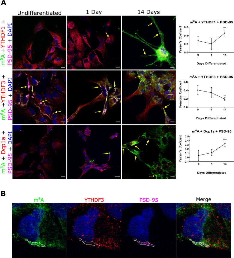
Fig. 4. m6A modified RNAs increase, and colocalization with YTHDF3 decreases, at synapses during synaptic maturation.
A Human neural progenitor cells examined by confocal microscopy show m6A-modified RNAs (green) increase in abundance during synaptic maturation (1 and 14 days differentiation) and increase in co-localisation at postsynaptic sites (magenta) with YTHDF1 (red) and Dcp1a (red) but decreases with YTHDF3 (red). Arrows point to regions where there is high white fluorescence signal indicating high colonisation of modified RNA and effector/dcp1a proteins at synapses. Far right, Pearson’s Correlation Coefficients for colocalisation between m6A modified transcripts, m6A reader binding proteins, YTHDF1, YTHDF3 and Dcp1a, over the three time points. B High magnification images showing less co-localisation between YTHDF3 (red) and m6A modified RNAs (green) at postsynaptic regions (PSD-95, magenta) encircled in white at day 14. Error bars denote 95% CI. *p ≤ 0.05, **p ≤ 0.005, ***p ≤ 0.0005.

