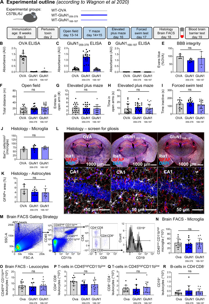
Fig. 5. Summary of the results from the replication study.
A Experimental outline, following the protocol of Wagnon et al. [17]. B–D Experimental validation of immunization success using OVA-ELISA, GluN1359–378-ELISA, and GluN1168-187-ELISA. E Blood–brain-barrier (BBB) integrity assessed through Evans blue extravasation. F–I Results of behavioral phenotyping, showing locomotor activity in the open field, anxiety-related behavior in elevated plus maze, and depression-like behavior in the forced-swim test. J–L Histological quantification using 8 mice/immunization with focus on reactive gliosis, showing microglia numbers, GFAP+ area (densitometry), and representative images of quantified stainings. High-resolution images of CA1 were acquired as 10 µm Z-stacks and displayed as maximum-intensity projections. M–R Characterization of the brains’ immune cell compartment by flow cytometry of 11–12 mice/group. M Gating strategy. Quantification of CD11bhighCD45mid cells (microglia). Quantification of CD11blowCD45high leukocytes, CD4+ T cells, CD8+ T cells, and CD19+ B cells. Data displayed as mean ±SD.

