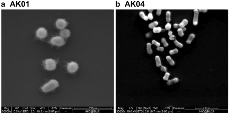
Fig. 1.
Scanning electron microscopy image of Arthrobacter sp. a strain AK01, b strain AK04. Cell morphology was examined using a scanning electron microscope (Quanta 200 ESEM). Cells were grown in LB media for 3 days, fixed in 0.05% glutaraldehyde in 0.1 M phosphate buffer (pH 7.4), then in 2.5% glutaraldehyde in 0.1 M phosphate buffer (pH 7.4) and allowed to react for 20 min. Fixed cells were adhered onto poly-lysine-coated slides and rinsed with water 3 times, then dehydrated by soaking in an ascending ethanol gradient (20–100%). The sample was critical point dried using a Leica CPD3000 and gold coated to thickness of 5 nm using Safematic CCU-010 compact coating unit. Images are at approximately 50,000 × magnification with scale bar shown

