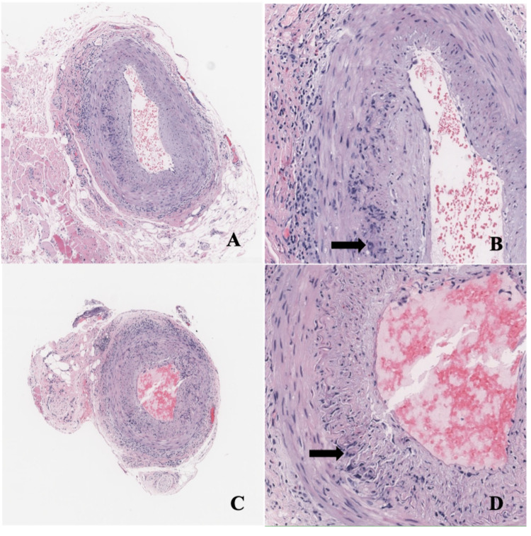
Figure 1. Temporal Artery Biopsies .
Right temporal artery (A, B) and left temporal artery (C, D): Medium size muscular artery with intramural inflammatory infiltrates composed of histiocytes, lymphocytes, and eosinophils. Multinucleated giant cells can be identified (arrows). The infiltrate is concentrated at the level of internal elastic lamina and adventitia. Hematoxylin and eosin (H&E) stain, magnification 50x (A, C), 200x (B, D).

