Figure 1.
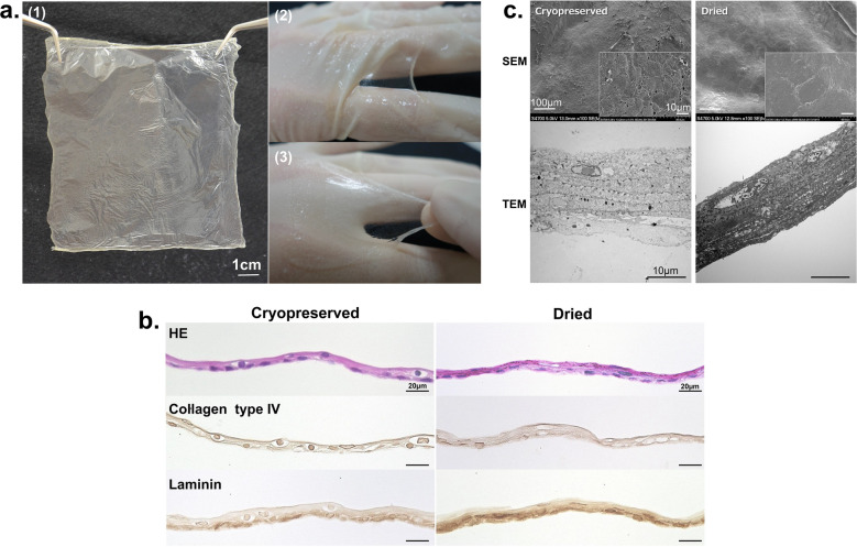
Morphological aspects of a dried CE. (a) Dried CE before hydration was cloudy, translucent, and fragile (1). The dried CE transformed into a transparent and flexible sheet with features similar to those of fresh CE sheets after rehydration (2,3). (b) Cryopreserved and dried CE sections stained with hematoxylin–eosin and anti-collagen type IV or anti-laminin antibodies. Both types of CEs exhibited similar morphological features, consisting of 4–5 layers of keratinocytes, including a monolayer of basal cells, constituting an undamaged membrane structure. The basal layers of both CEs were detected using immunostaining with an anti-laminin antibody, whereas the collagen type IV staining was not clear. (c) SEM and TEM images of cryopreserved and dried CEs. (TEM) Five to six layers of keratinocytes, including the monolayer of basal cell and desmosomal complexes at cell contact points, were observed in both CEs. (SEM) Both CEs appeared similar; the keratinocytes formed a smooth and regular membrane. No crack was observed on the membrane surface. CE, cultured epidermis; SEM, scanning electron microscopy; TEM, transmission electron microscopy.

