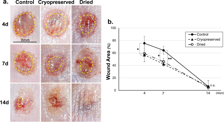
Figure 4.
Wounds on the skin defect created on the back of diabetic mice. (a) Yellow lines indicate the wound area. In the cryopreserved CE and dried CE groups, no wounds remained on the representative samples on day 14. (b) The wound areas in the cryopreserved CE, dried CE, and control groups on days 4, 7, and 14 are depicted. Four and seven days after the operation, the wound areas in the two CE groups were significantly smaller than that in the control group (*P < 0.05, **P < 0.01, n = 6). The wound almost closed in all groups 14 days after the operation. CE, cultured epidermis.

