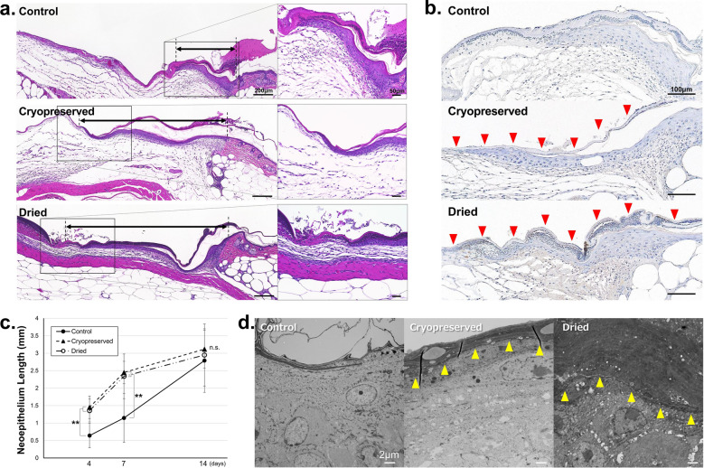
Figure 5.
Microscopic images of wound areas in mice of control, cryopreserved CE, and dried CE groups. (a) The hematoxylin–eosin stained sections of the wounds in control, cryopreserved CE, and dried CE groups. The arrows indicate the newly formed epithelium. (b) Sections stained with anti-STEM121 in the wounds of control, cryopreserved CE, and dried CE groups. The red arrowheads (filled triangle) indicate cultured epidermis. The recipient murine keratinocytes can be easily distinguished from the transplanted CE. (c) Length of the epithelium in the wounds in control, cryopreserved CE, and dried CE groups. The epithelial lengths of the two CE groups were significantly higher than those of the control group on days 4 and 7, and significant difference was not observed between the two CE groups. Significant differences between the groups were not observed on day 14. (**P < 0.01, n = 6). (d) Transmission electron microscopy (TEM) images of the wound edge in control, cryopreserved CE, and dried CE groups. The wounds were harvested on day 2 and observed using TEM. The yellow arrowheads (filled triangle) indicate the boundary between the implanted CE and recipient keratinocytes. The transplanted human CE and the cell membrane of the mouse keratinocytes completely adhered without any gaps. CE, cryopreserved epidermis.

