Fig. 1.
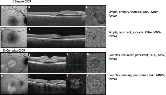
i) Representative cases showing simple central serous chorioretinopathy (CSCR).First panel: A 38-year-old patient presented with metamorphopsia for 3 weeks in their right eye, best corrected visual acuity (BCVA) of 20/20 (Snellen) and no history of recurrence. A Fundus autofluorescence (FAF) image demonstrating < 2-disc diameters (DD) of retinal pigment epithelium (RPE) alteration thereby classifying the case as simple CSCR. B Optical coherence tomography (OCT) B scan passing in the horizontal direction through the fovea demonstrating subretinal fluid (SRF) and no evidence of outer retinal atrophy (ORA). C OCT angiography (OCTA) at the level of choriocapillaris is unremarkable. The eye is classified as Simple CSCR, primary episode, sporadic (<6 months duration), absent ORA (ORA-), with foveal involvement (fovea + ) and absent macular neovascularization (MNV-). Second panel: A 44-year-old patient presented with metamorphopsia for 2 months in their left eye, BCVA of 20/15 (Snellen) and a history of previous such episode. D FAF imaging demonstrates < 2 DD of RPE alteration, E OCT B scan passing in the horizontal direction through the fovea shows SRF, a tiny pigment epithelial detachment (PED) and no evidence of ORA (ORA-). F OCTA at the level of choriocapillaris is unremarkable. The eye is classified as Simple CSCR, recurrent episode, sporadic, ORA-, fovea + , MNV-. 1 ii). Representative cases showing complex central serous chorioretinopathy (CSCR). Third panel: A 38-year-old patient presented with complaints of blurred vision for 6.5 months in their right eye with a history of previous such episode. Best corrected visual acuity (BCVA) was 20/20. A Fundus autofluorescence (FAF) image demonstrating > 2-disc diameters (DD) of retinal pigment epithelium (RPE) alteration (multifocal) thereby classifying the case as complex CSCR. B Optical coherence tomography (OCT) B scan passing in the horizontal direction through the fovea demonstrating subretinal fluid (SRF) and no evidence of outer retinal atrophy (ORA -). OCT angiography (OCTA) at the level of outer retina (C) and choriocapillaris (D) is unremarkable. The eye is classified as complex CSCR, recurrent episode, persistent (>6 months duration), ORA-, with foveal involvement (fovea + ) and absent macular neovascularization (MNV-). Fourth panel: A 49-year-old patient complained of decreased vision in right eye for 2 years. BCVA was 20/200 with no history of previous such episode. E FAF imaging shows > 2 DD of RPE alteration thereby classifying the case as complex CSCR. F OCT B scan passing in the horizontal direction through the fovea demonstrating SRF and flat irregular pigment epithelial detachment (FIPED). OCTA at the level of outer retina (G) and choriocapillaris (H) shows MNV (MNV + ). Thus, the eye was classified as complex CSCR, primary episode, persistent SRF, ORA + , fovea + , MNV + .

