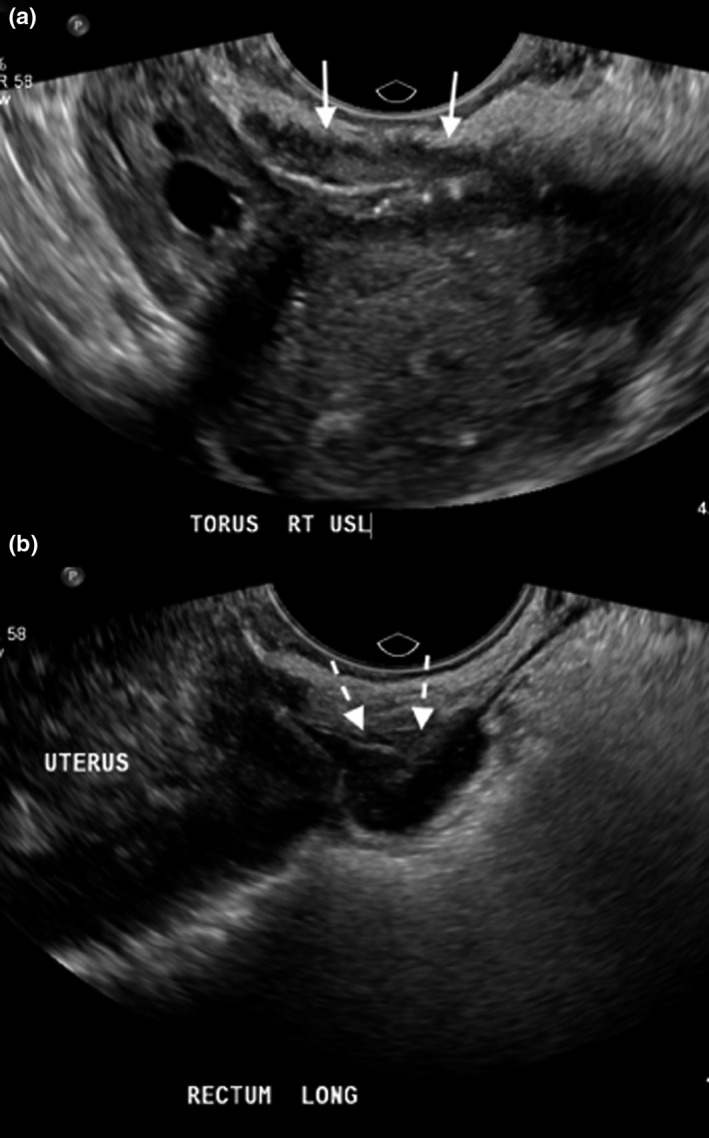
Figure 3.

Two nodules of endometriosis seen within the pelvis of the case which took 42 minutes. A large nodule of endometriosis is seen within the right uterosacral ligament, extending into the torus uterinus as shown by the white solid arrows (a). A nodule of endometriosis was also seen within the rectal wall (b) as depicted by the dashed arrows. Also present in this case were extensive pelvic adhesions, including obliteration of the pouch of Douglas and a left ovarian cyst (not pictured).
