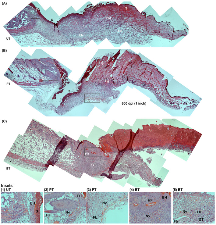
FIGURE 7.

Progression of healing at 3 dPIT. Tissue from UT, PT, and BT wounds were H&E stained and examined at ×100 for evidence of inflammatory and proliferative stages of healing. Images were obtained and processed as described for Figure 4. The representative panoramic images show half of the wound bed plus normal skin at the wound margin. Insets indicated by black boxes in panoramas were taken at ×200 and are numbered. A, UT wound with inset 1 showing epidermal hyperplasia; B, PT wound with insets 2 and 3 showing epidermal hyperplasia, regenerating hair follicle, and neovascularisation; C, BT wound with insets 4 and 5. BT, BDWG‐treated; Ed, edema; EH, epidermal hyperplasia; Fb, fibroblasts; fS, forming scab; GT, granulation tissue; H&E, haematoxylin and eosin; HF, hair follicle; Nv, neovascularisation; PM, provisional matrix; R, regeneration of fat cells; S, scab; PT, PEG‐treated; PEG, polyethylene glycol; UT, untreated
