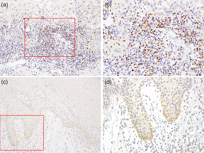
Figure 1.

FoxP3 and HPV were detected using immunohistochemistry. (a, b) FoxP3 was stained in the nuclei of cells that were believed to be Tregs infiltrating the peritumoral stroma. (c, d) HPV antigens were stained in cells forming the base membrane of cancer tissues. Magnification 200× (a, c); 400× (b, d)
