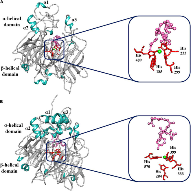
FIGURE 6.
Best-fit 3D model and docking analysis of (A) BoCCD1-4 and (B) BoCCD4-2 proteins. The α-helix (α1, α2, and α3) and the β-propeller domains are indicated on the models. Green shows the Fe2+ ion coordinated by the four histidine residues. The four histidine residues are shown in red, and the lycopene molecule is shown in pink.

