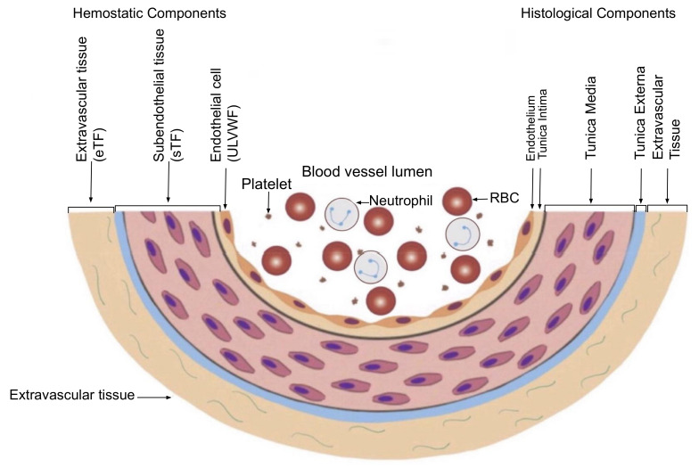
Figure 3.
Schematic illustration of cross section of blood vessel histology and hemostatic components (Reproduced and modified with permission from Chang JC. Clin Appl Thromb Hemost 2019 Jan–Dec; 25:1076029619887437) [11]. The blood vessel wall is the site of hemostasis (coagulation) to produce blood clots (hemostatic plug) and to stop hemorrhage in external bodily injury. It is also the site of hemostasis (thrombogenesis) to produce intravascular blood clot (thrombus) in intravascular injury that causes thrombosis. Its histologic components are divided into the endothelium, tunica intima, tunica media and tunica externa, and each component has its function contributing to molecular hemostasis. As illustrated, ECs damage triggers exocytosis of ULVWF and SET damage promotes the release of sTF from tunica intima, tunica media and tunica externa. EVT damage releases of eTF from the outside of blood vessel wall. This depth of blood vessel injury contributes to the genesis of different thrombotic disorders such as microthrombosis, macrothrombosis, fibrin clots/hematoma and thrombo-hemorrhagic clots. This concept is especially important in the understanding of different phenotypes of stroke and heart attack. Abbreviations: EVT, extravascular tissue; eTF, extravascular tissue factor; SET, subendothelial tissue; sTF, subendothelial tissue factor; RBC, red blood cells; ULVWF, ultra large von Willebrand factor.

