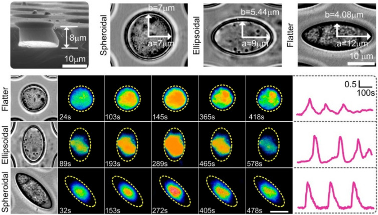
Figure 4.
Bright field images of chondrocytes show single cell encapsulated in a microniche with different geometries. Representative single cell images showing Ca2+ oscillations at given time points from one chondrocyte in the spheroidala, ellipsoidala, and flattera microniches. Yellow dashed line represents the boundaries of microniche. TRPV4 and PIEZOs may be involved in mediating calcium signaling. Figure based on our recent publication [67].

