Figure 3.
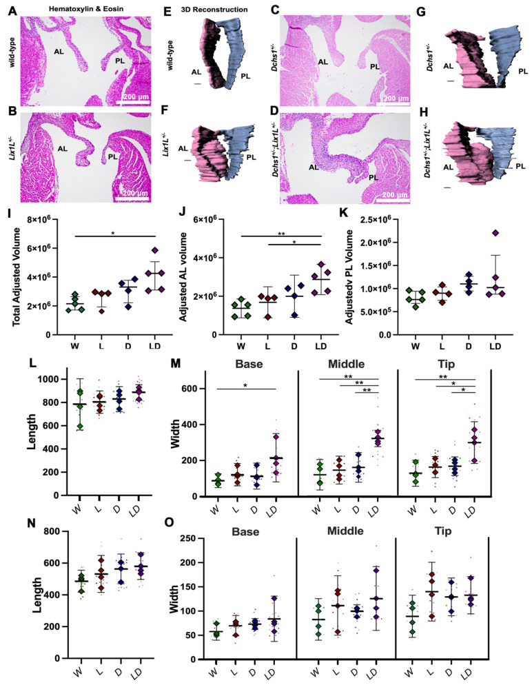
Epistasis analysis reveals DCHS1-LIX1L genetic interaction at postnatal day zero. (A–D) Hematoxylin and eosin (H&E) staining and (E–H) 3D reconstructions of postnatal day zero (P0) mitral valves isolated from wildtype (W), single heterozygote (LIX1L+/−, L or DCHS1+/−, D) and compound heterozygote (LIX1L+/−;DCHS1+/−, LD) mice reveal thickening of single heterozygote and compound heterozygote anterior leaflets (AL) compared to wildtype control littermates. (I–K) Total mitral valve volume measurements from 3D-reconstructed leaflet surfaces demonstrate a significant increase in compound heterozygotes AL leaflets compared to wildtype, with no significance observed in posterior leaflet (PL) volumes. Two-dimensional measurements of length (L,N) and width (M,O) along the leaflet reveals that changes in leaflet volume are due to significant leaflet thickening in the base and tip of the (L,M) anterior leaflets of compound heterozygotes versus single heterozygotes or wildtypes. (N,O) No significant differences were observed in the posterior leaflet of these mice. N = 4–5 animals per genotype, 2D measurements completed on four sections per animal and depicted by dots, averages are plotted as diamonds, * p < 0.05, ** p < 0.005 with One-Way Anova.

