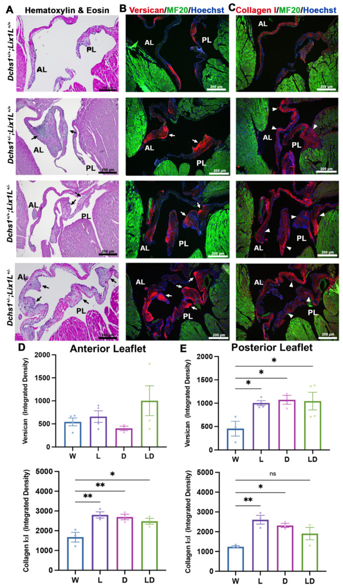
Figure 4.
Adult heterozygotes display mitral valve disease phenotype of myxomatous degeneration. (A) Representative H&E staining of 11-month-old wildtype (W), single (L, D) and compound heterozygote (LD) mitral valves depict thickening and bulging of single and compound heterozygotes anterior and posterior leaflets (AL, PL) (black arrows). Immunohistochemical (IHC) staining of (B) Versican (red) and (C) Collagen 1α1 (red), myocardium (MF20, green) and nuclei (Hoechst, blue) reveals increased extracellular matrix deposition and disorganization (white arrows and arrowheads) in single or compound heterozygotes. (D,E) Quantification of ECM staining intensity reveals statistically significant increases in single or compound heterozygote leaflets compared to wildtype controls, indications of myxomatous degeneration. N = 3 per genotype, depicted with mean and SEM, * p < 0.05, ** p < 0.005 with One Way Anova.

