Figure 5.
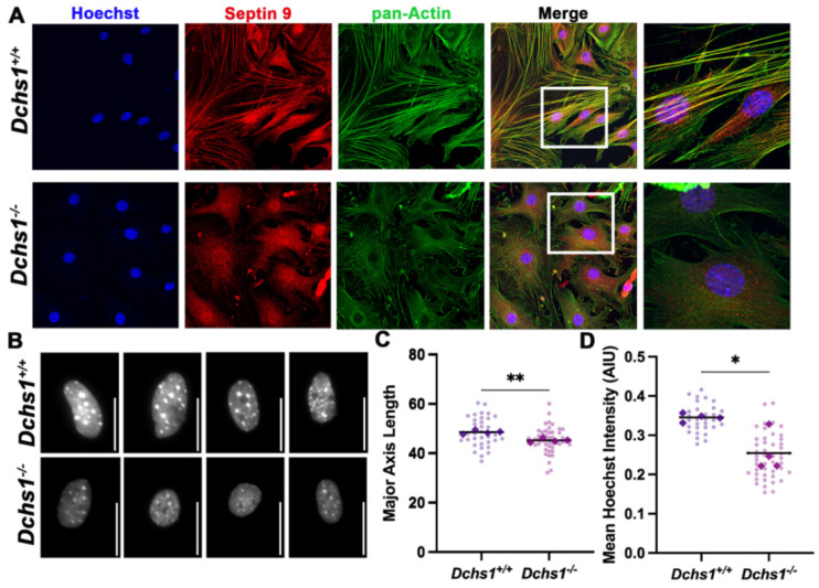
Septin-actin defects in DCHS1-deficient fibroblasts. (A) Wildtype (DCHS1+/+) and knock-out, (DCHS1−/−), cardiac fibroblasts (CFs) were seeded on collagen coated slides and stained for pan-actin (green), septin 9 (red) and nuclei (blue). In DCHS1−/− fibroblasts, actin and SEPT9 no longer co-localize coincident with loss in stress fiber organization and altered cell shape. (B) Representative grayscale images of nuclei stained with Hoechst illustrate a rounded phenotype in DCHS-−/−, scale bar = 50 pixels. (C) Quantification of nuclei major axis length (pixels) and (D) Hoechst staining intensity (Arbitrary intensity units, AIU) reveals significant reductions in knockout cells compared to wildtype controls suggesting loss of intracellular tension. 30–50 cells imaged per genotype, N = 4 ** p < 0.005, * p < 0.05 with Student’s t-test.

