Figure 7.
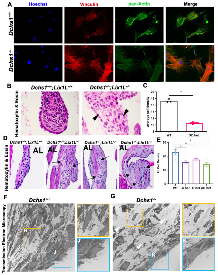
Consequences of actin defects. (A) ICC of vinculin (red), pan-actin (green), and nuclei (Hoechst, blue) in wildtype (DCHS1+/+) and DCHS1 KO (DCHS1−/−) reveals loss of stress fiber formation and vinculin organization and localization to focal adhesions. (B) H&E images of wildtype and compound heterozygote P0 valves depict increased density of ECM between interstitial cells (black arrowheads), also observed in (D) 11-month-old leaflets of single and compound heterozygote anterior leaflets (arrows). (C,E) Quantification of cell density measured by cells divided by total leaflet area reveals significant decreases in compound heterozygotes (XD het) compared to controls at P0 and both single and compound heterozygotes in adults. Graphs depict average cell density per animal with error bars of SEM, n = 3–4 animals per genotype, * p < 0.005. (F,G) Transmission electron microscopy (TEM) images of wildtype and DCHS1 KO anterior leaflets at P0 show increased ECM content in DCHS1 KOs and less dense interstitium with round nuclei and loss of filopodia-structures (yellow boxes, (H,I)). Filopodia-like structures appear to be lost along the DCHS1 KO endothelium (blue boxes, J,K).

