Figure 2.
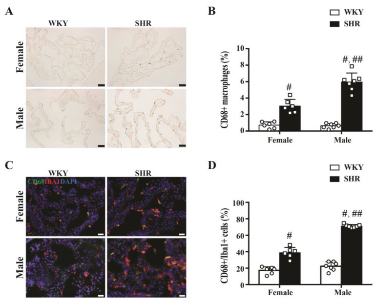
(A) Example of CD68 immunoreactivity in the choroid plexus of male and female SHRs and WKYs at week 9 (scale bar = 50 μm). (B) CD68-positive cells as a percentage of all choroid plexus cells in male and female SHRs and WKYs at week 9. Values are the means ± SD, n = 6−7; # p < 0.01 compared with the WKY groups. Male and female SHRs had more CD68+ cells than WKYs. In addition, male SHRs had more CD68-positive cells than female SHRs; ## p < 0.01 compared with the female SHR group. (C) Co-localization of CD68- and Iba1-positive cells at the choroid plexus in male and female rats at week 9 (scale bar = 20 μm). (D) Percentage of Iba-positive cells that were also positive for CD68 at the choroid plexus. Values are the means ± SD, n = 6–7; # p < 0.01compared with the WKY groups. In both male and female SHRs, a greater % of Iba+ cells were CD68+ than in WKYs. In addition, male SHRs had a greater % than female SHRs; ## p < 0.01 compared with the female SHR group.

