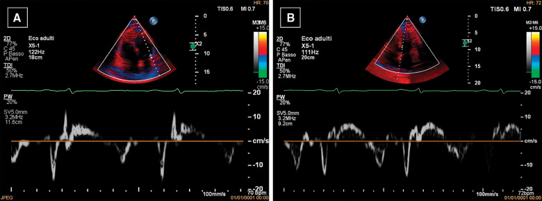
Figure 2.
Echocardiography. Lateral pulsed wave tissue Doppler imaging (PW-TDI) e′ values (9 cm/s) (A) are lower than septal PW-TDI e′ values (11 cm/s) (B), as opposed to normal. This finding is known as ‘annulus paradoxus’ and is caused by tethering of the lateral wall by the thickened pericardium.

