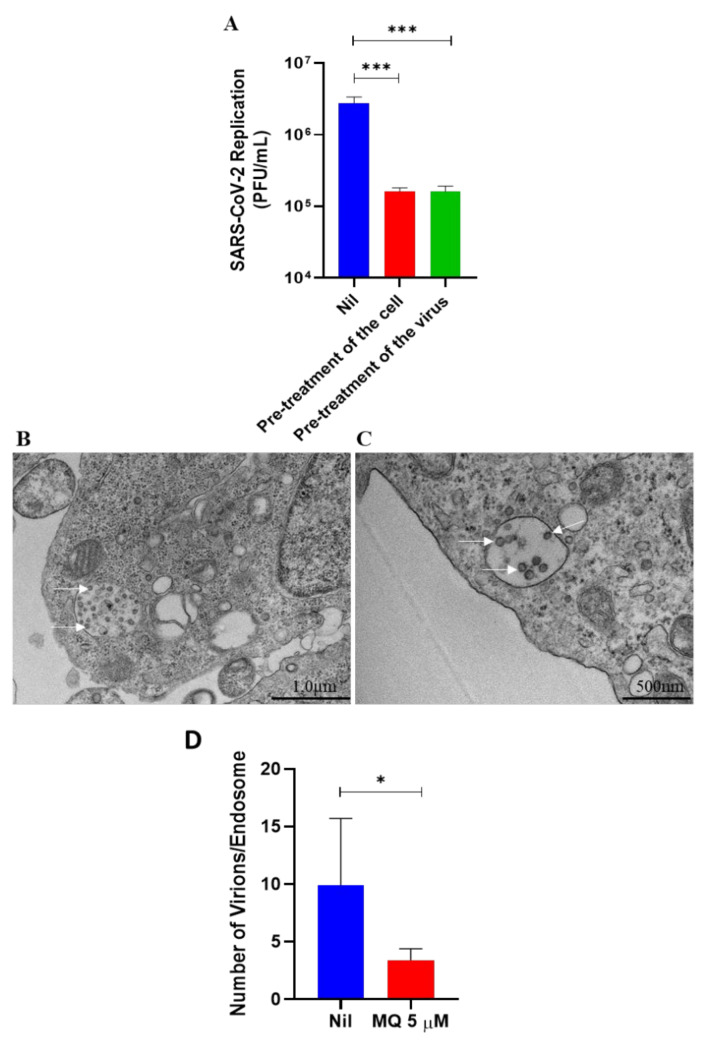
Figure 4.
Effects of mefloquine on SARS-CoV-2 endocytosis-mediated entry. (A) To initially evaluate mefloquine’s effect on virus entry, Calu-3 cells or SARS-CoV-2 virus particles were pre-incubated with 1 µM of mefloquine for 1 h at 37 °C and then infected at an MOI of 0.1. After 24 hpi, culture supernatants were harvested, and SARS-CoV-2 replication was measured using the plaque assay. Results are displayed as virus titers (PFU/mL). (B,C) Representative images (from 4 independent experiments) of ultrastructural analysis by transmission electron microscopy of Vero E6 cell infected with an MOI of 1 of SARS-CoV-2 (B) and treated with 5 µM of mefloquine for 4 h (C). Cell endosomes with spherical SARS-CoV-2 virus particles (white arrows). (D) SARS-CoV-2 virus particles were counted inside the endosomes of Vero E6 cells treated with mefloquine or not (nil). * p < 0.05 and *** p < 0.01.

