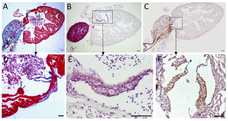
Figure 3.
Elastin content of the zebrafish heart. Histological sections of adult zebrafish entire hearts (A–C) and cardiac valves (D–F). Sections were stained following different protocols: Masson-Goldner trichrome staining (A,D) makes muscle fibers appear in red, collagen in blue, and erythrocytes in pinkish violet; orcein (B,E) stains elastin in pink and nuclei appear in violet due to hematoxylin staining; immunohistochemistry against elastin (C,F) with elastin signal appearing in brown; (D,E) are atrioventricular valves, (F) shows the bulboventricular valve. Scale bars: 100 µm (A–C); 50 µm (D–F). Magnification: 40× (A–C); 200× (D,F); 00× (E).

