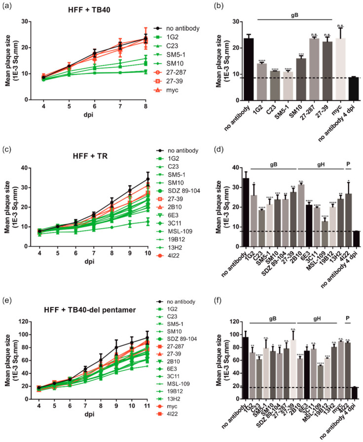
Figure 3.

Anti-gH and -gB antibodies limit HCMV spread in fibroblasts in a strain-independent manner. HFFs in 96-well plates were infected with 100 PFU/well of different HCMV strains ((a,b) TB40/E; (c,d) TB40/E-del pentamer; (e,f) TR). After incubation for 24 h, the medium was replaced by agarose overlay medium with or without the indicated antibodies (50 µg/mL). Starting at 4 dpi, whole 96-well images were captured each day for at least up to 19 dpi and used for automated quantification of the mean plaque size (1E-3 Sq.mm) of all individual fluorescent spots detected per well as described in Materials and Methods. All experiments were performed in triplicate. (a,c,e) Time course analyses of the mean plaque sizes of HFF cells infected with the HCMV strains TB40/E (a), TB40/E-del pentamer (e), or TR (c). nt mAbs are colored in green, nnt and negative control mAbs are shown in red. (b,d,f) Bar graphs of the mean plaque sizes at the time points post-infection with maximum mean plaque sizes of the no antibody controls ((b), 9 dpi; (d), 10 dpi; (f), 11 dpi). The dashed lines indicate the mean plaque sizes of the no antibody controls at 4 dpi, which is indicative of the initial fluorescent spot size of a single-round infection. Statistical analysis was performed by ordinary one-way analysis of variance (ANOVA). n.s., not significant; *, p ≤ 0.1; **, p ≤ 0.01; ***, p ≤ 0.001; ****, p ≤ 0.0001; p values refer to antibodies vs. no antibody control.
