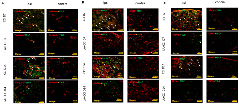
Figure 3.
LA decreased not only expression of small-sized but also expression of large-sized DRG neurons, esp. nociceptive DRG neurons. The distribution of TSLP was detected when DRG neurons were double-labeled (yellow) with TSLP (green) and neuronal markers (red). LA serves as a TSLP inhibitor. (A) LA decreased the TSLP-positive small-sized DRG neurons, (B) TSLP-positive large-sized DRG neurons, and (C) TSLP-positive nociceptive DRG neurons in the ipsilateral (ipsi) injured side as compared to that in the contralateral (contra) side. Pairs of merged images are shown in the right panels. White arrows indicate doubled-labeled cells. Scale bars represent 50 μm (n = 6 rats).

