Figure 5.
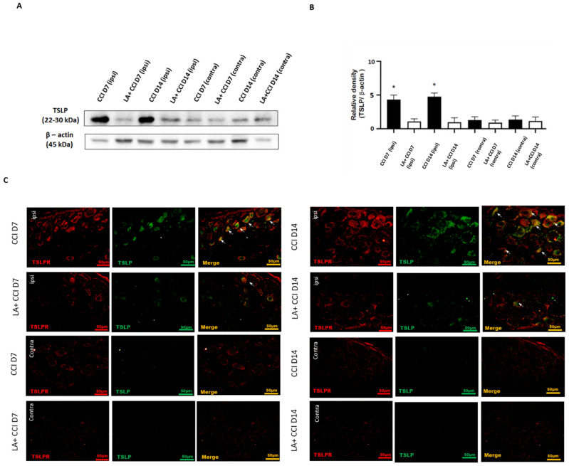
LA attenuated the mechanical hyperalgesia through the TSLP/TSLPR complex. (A) Lumbar 4/5th DRG at both sides were dissected. TSLP proteins were measured by western blot analysis when β-actin was used as the loading control. At day 7 and day 14 after CCI, TSLP proteins were increased in DRG in the ipsilateral injured side as compared to those in the contralateral side. LA reduced the TSLP expression in the ipsilateral injured side. (B) Each band signal density was quantitated, and normalized to that of its own β-actin in each side. Values are presented as means ± s.e.m. (n = 6 rats). * p < 0.05, compared to LA administration group, Students’ t test. (C) The distribution of TSLP was detected when DRG neurons were double-labeled (yellow) with TSLP (green) and TSLPR (red). Merge immunoreactivities were parallel-measured in the ipsilateral and contralateral sides of DRG neurons at 7 and 14 days. This expression of TSLP/TSLPR in the ipsilateral (ipsi) injured side increased after nerve injury, and decreased LA administration as compared to that in the contralateral (contra) side. Pairs of merged images are shown in the right panels. White arrows indicate doubled-labeled cells. Scale bars represent 50 μm (n = 6 rats).

