
Figure 4.
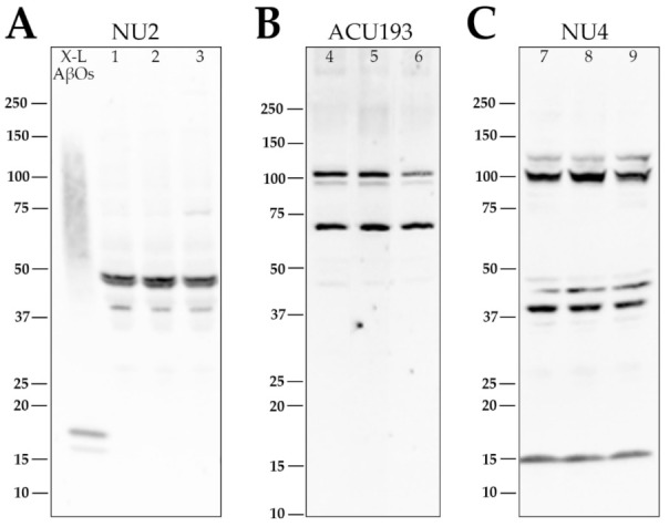
Retina AβOs comprise proteoforms differentially recognized on Western blots by monoclonal antibodies NU2, ACU193, and NU4. Soluble extracts from three embryonic retinas were obtained at E14 and separated using SDS-PAGE (Tris-glycine gel) followed by transfer to a nitrocellulose membrane. This was repeated twice for a total of nine different retinas (1–9) on three separate membranes. AβOs were identified using NU2 (A; 1–3), ACU193 (B; 4–6), or NU4 (C; 7–9). AβOs identified with NU2 were most prominent as a doublet at ~45 kDa with a minor band just above 37 kDa. AβOs identified with ACU193 had prominent bands at ~72 kDa and a doublet at ~100 kDa. AβOs identified with NU4 had prominent bands just above ~37 kDa and ~100 kDa, with fainter bands at ~14 kDa, 45 kDa, and 125 kDa. The ability of NU2, ACU193, and NU4 to distinguish these distinct SDS-stable AβO proteoforms was observed in three separate experiments.