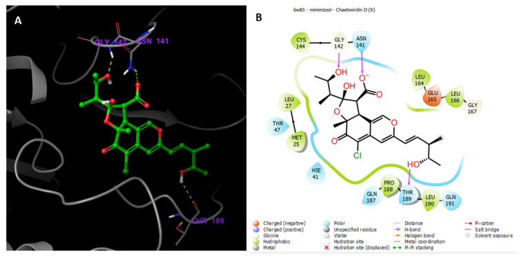
Figure 10.
(A) Putative binding mode of chaetovirdin D in the binding site of 2019-nCOV 3CL hydrolase (Mpro), PDB: 6W81. Chaetovirdin D is displayed as green sticks. The amino acids of the binding site are represented as grey sticks, and H-bonds are represented as yellow dotted lines. (B) 2D depiction of the ligand–protein interactions.

