Figure 7.

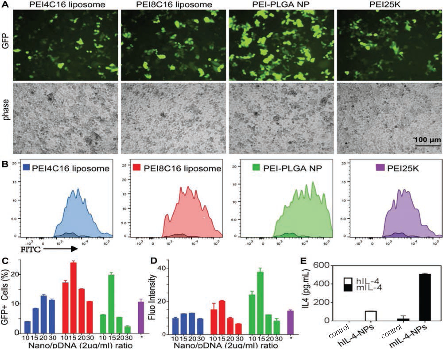
Fluorescence microscopy and flow cytometry assays were used to determine the transfection of pIL-4/GFP loaded PEI4C16 liposome, PEI8C16 liposome, PEI-PLGA NP and PEI25K under their best transfection conditions. The transfection experiments were performed with pIL-4/GFP concentration at 4 µg/mL in PEI4C16 liposome, and 2 µg/mL in PEI8C16 liposome and PEI-PLGA NP. The transfection by PEI25K was at its optimized N/P ratio of 10/1 (4 µg/mL). The images of GFP expression were observed at various N/P ratios (A). Transfection efficiency of pIL-4/GFP in cells was determined by flow cytometry (B). Quantitative analysis of fluorescence microscopy images was used to calculate the percentage of GFP positive cells (C). The levels of GFP expression were measured by flow cytometry to determine the fluorescence intensity (D). To identify the expression of exogenous IL4, mouse bone marrow derived macrophages (BMMs) were transfected with human pIL-4 (hIL-4) and mouse pIL-4 (mIL-4) (E). The culture medium were collected to perform ELISA test. The levels of human and mouse IL-4 protein in BMMs culture medium were significantly increased at day 5 post-transfection as compared to the control groups (E). Data are expressed as mean ± SD (n = 3) (P < 0.01).
