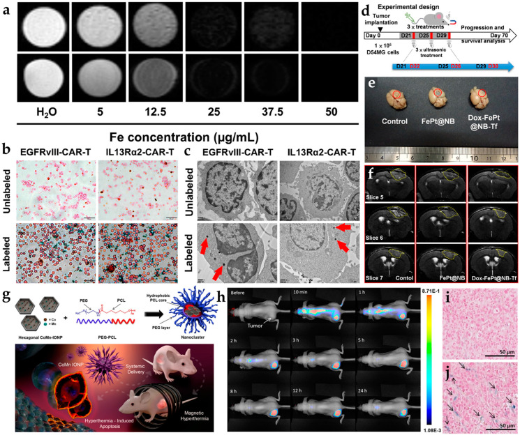
Figure 3.
The various types of iron-based nanoplatforms in glioblastoma theranostics. (a) T2-weighted images containing iron-oxide nanoparticles labeled CAR-T cells, EGFRvIII CAR-T (upper row), and IL13Rα2 CAR-T (lower row) cells. (b) There are representative images of Prussian-blue-stained, unlabeled CAR-T cells (top) and labeled CAR-T cells (bottom). (c) Cell TEM views of unlabeled CAR-T cells (top) and labeled CAR-T cells (bottom) [18]. (d) Illustrative mice model of synthesis and HIFU-triggered drug release from FePt@NB. (e) Photos of mouse brains with GBMs. The GBM tumors are circled with a yellow highlight. (f) T2-weighted MRI images of mouse brains with GBMs. The GBM tumors are highlighted in yellow circles [32]. (g) Graphical representation of CoMn IONP encapsulated inside PEG/PCL polymers. (h) IVIS system analyzed for injection of CoMn-IONP nanoclusters loaded with a hydrophobic NIR dye. Prussian-blue staining of tumor slices of (i) 5% dextrose and (j) CoMn-IONP nanoclusters [34]. Adapted with permission from Refs. [18,32,34], Copyright 2021 Elsevier, 2019 and 2021 American Chemical Society.

