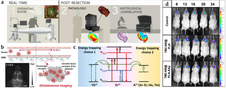
Figure 4.
The common nanoprobes in glioblastoma imaging. (a) Three kinds of workflow in glioblastoma imaging with cetuximab-IRDye800D [48]. (b) Illustration of blood–brain barrier crossing with targeted NIR-II fluorescence in brain vessel imaging [64]. (c) Mechanism of energy transfer with Yb3+, Er3+, and A3+ downconversion system [69]. (d) The IVIS image from persistent luminescent ZnGa2O4:Cr3+, Sn4+ with the tumor change (days 8, 12, 16, 20, and 24) [75]. Adapted with permission from Refs. [48,64,69,75]. Copyright 2018 Springer, 2020 Wiley, 2020 Elsevier and 2021 American Chemical Society.

