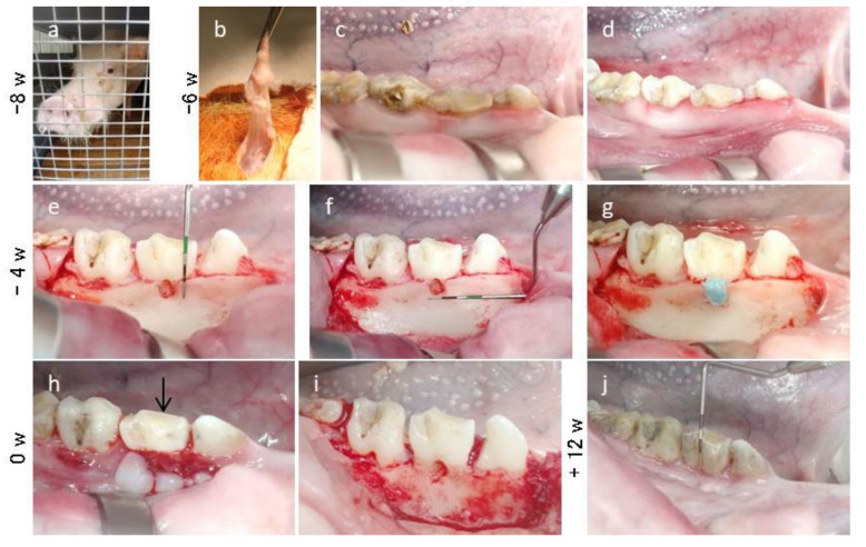
Figure 3.
Creation of the periodontal furcation defect in the mandible of MMPs. (a) MMP at the Nihon University School of Medicine. (b) Small pieces of adipose tissue obtained from the hypogastrium. (c,d) Removal of the calculus in the supragingival region of mandible premolars using a curette. (e,f) The furcation defects (4 mm wide, 5 mm deep, and 3 mm horizontally) were created on the buccal side of the bilateral second premolars. (g) Filling the impression material into the created furcation defect to induce chronic inflammation. (h) Four weeks later, inflammation was observed at the buccal surface. Black arrow indicates gingival inflammation and bleeding from the buccal side of the bilateral second premolar. (i) After debridement, the furcation defect and the buccal bone of premolars exhibited destruction. (j) Twelve weeks later, the clinical parameters were once again measured before extracting the mandible.

