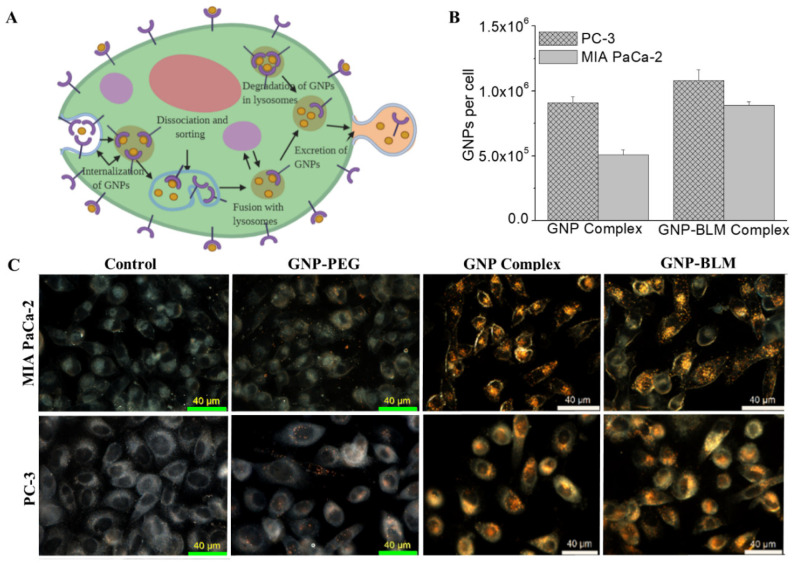
Figure 3.
Cellular uptake of GNP complexes. (A) endo-lysosomal path of NPs within a cell. (B) Cellular uptake comparison of GNP-BLM and GNP. (C) Darkfield (scale bar = 40 μm) imaging of control cells and cells internalized with GNPs, GNP–PEG, GNP complex (GNP-PEG-RGD) and GNP-BLM (GNP-PEG-RGD-BLM). Yellow bright spots in darkfield images indicates the presence of NP clusters.

