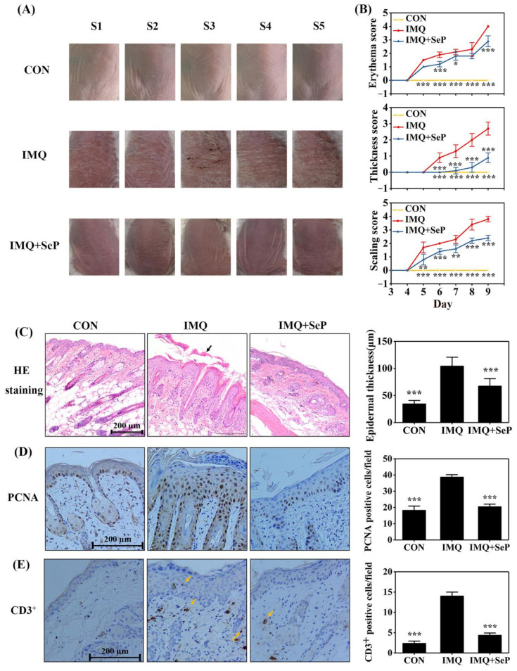
Figure 1.
Effect of SeP on the IMQ-induced psoriasis-like dermatitis in mice. (A) The macroscopic appearance of mice back skin on day 9. The back skin of 5 mice (S1–5) were shown in each group. (B) The symptoms of erythema, skin thickness, and scaling were scored on day 3 to day 9 based on the PASI. (C) Hematoxylin-eosin (HE) staining of dorsal skin on day 9 (left panel) and quantification of the epidermal thickness (right panel). The black arrow indicated the sloughed scales. (D,E) Immunohistochemical staining for PCNA (D) and CD3 (E) in mouse dorsal skin on day 9. Representative images were shown in left panel and quantification of PCNA or CD3+ positive cells were shown in right panel. Quantification of the epidermal thickness, PCNA-positive cells and CD3+ positive cells of mouse back skin were obtained from 5 to 8 sites per mouse per group. Data were expressed as mean ± SD (n = 6). * p < 0.05, ** p < 0.01, *** p < 0.001, compared with the IMQ group.

