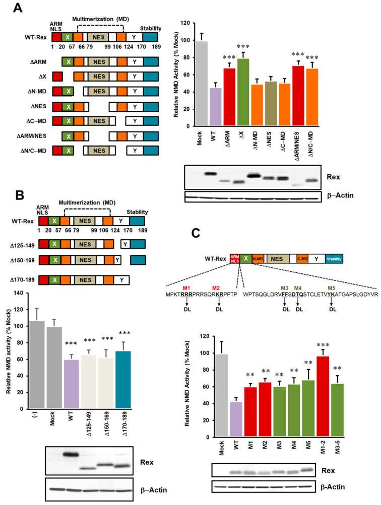
Figure 2.
N-terminal region of Rex plays important roles in NMD inhibition. (A). We generated seven domain-deficient mutants of Rex (left panel) and compared the degree of NMD repression with WT-Rex using the NMD activity reporter assay system in HeLa cells. As shown in the graph on the right, NMD repression was significantly reduced when the ARM region, X region or both multimerization domains were deleted. Western blotting below shows the expression levels of each Rex mutant (n = 6, mean ± SD, *** p < 0.001, compared with WT-Rex). (B). For the C-terminal side, where the stabilizing domain of Rex is located, we also generated three types of deletion mutants (upper panel) and compared the degree of NMD repression with WT-Rex using the NMD activity reporter assay system in HeLa cells. As shown in the lower graph, there was no significant change in NMD repression in any of the mutants compared to WT-Rex. Western blotting below shows the expression levels of each Rex mutant (n = 6, mean ± SD, *** p < 0.001, compared with Mock). (C). We generated five point mutants in the ARM and X regions of Rex (upper panel) and compared the degree of NMD repression with WT-Rex using the NMD activity reporter assay system in HeLa cells. As shown in the lower graph, the degree of NMD suppression was significantly reduced in all mutants compared to WT-Rex. The reduction in NMD repression was particularly pronounced for both M1 and M2 mutants. Western blotting below shows the expression levels of each Rex mutant (n = 6, mean ± SD, ** p < 0.01; *** p < 0.001, compared with WT-Rex).

